Anatomy of the thoracic cage and the breast: illustrations

Anatomy of the thoracic cage and the breast: illustrations

Anatomy of the thoracic cage and the breast: illustrations


This e-Anatomy module presents labeled illustrations of the thoracic wall and breast in 64 anatomical drawings with over 260 anatomical structures.


Breast - Anatomy : Sagittal section (Illustrations: A. Micheau - MD - Imaios)


Useful tool for learning and teaching anatomy, especially for students of medicine or paramedical health disciplines (physiotherapists, nurses, radiology manipulators, osteopaths...).

Anatomical illustrations of the thoracic cage and the mammary gland

The illustrations were drawn in Adobe Illustrator using data from medical imaging (principally CT scanner with 3D reconstructions).
They represent diagrams for the study of the anatomy of the thoracic wall and breast:

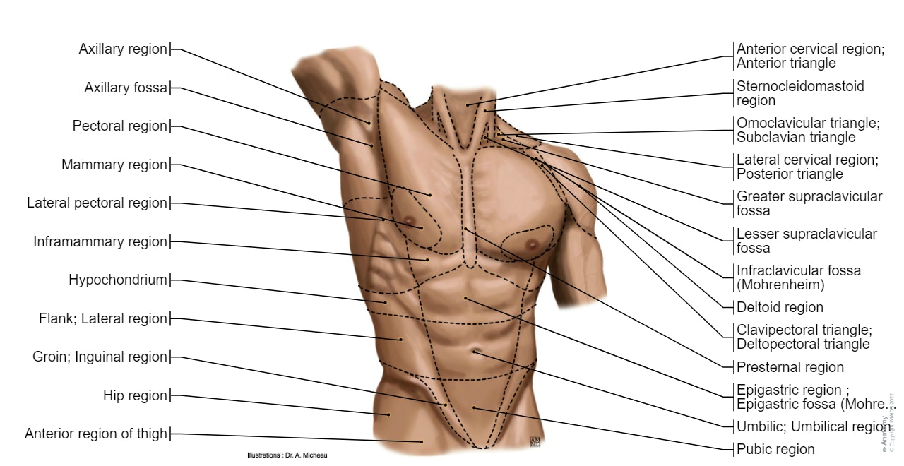
  • Surface anatomy: illustrations in anterior and posterior view of male torso and back, allowing the lines and regions used in surface anatomy to be displayed (midclavicular line, midline, pectoral region, sternal region...)


Trunk - Surface anatomy : Pectoral region, Mammary region, Hypochondrium, Presternal region, Midclavicular line


  • Thoracic skeleton: patterns of bony anatomy of the thoracic cavity and rib cage in anterior and posterior view.
Anatomy of the thoracic wall and the breast (Illustrations) : Thoracic cage - Thoracic cavity


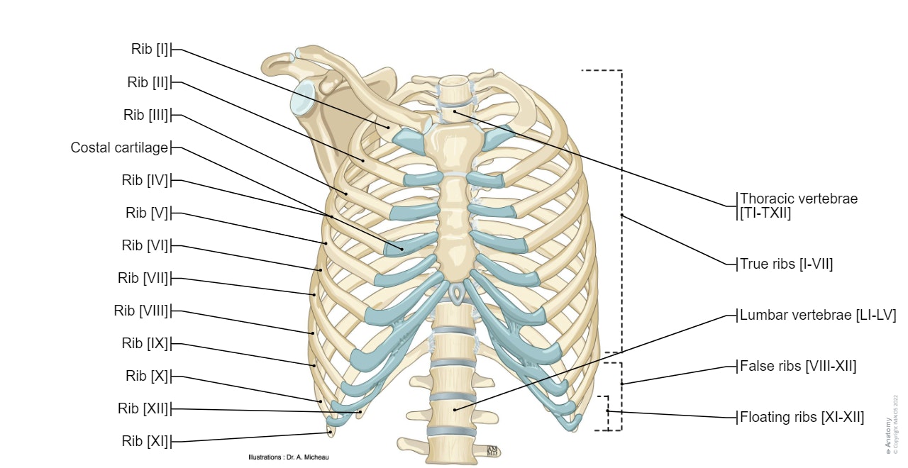
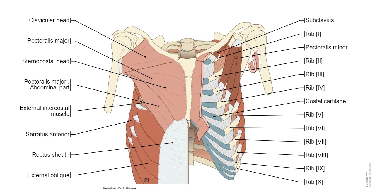
  • Ribs: represents the anatomy of the ribs and muscle attachments
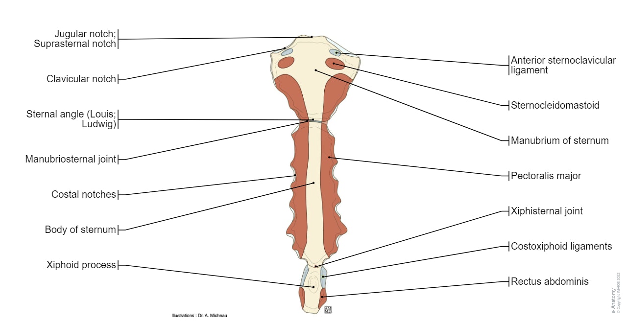
  • Sternum: details the different parts of the sternum (manubrium, sternal angle, xiphisternal joint) and the different muscle insertions (pectoralis major and sternocleidomastoid muscles).
Sternum (Muscles-Attachment) : Manubrium of sternum Clavicular notch Jugular notch; Suprasternal notch Sternal angle Body of sternum Xiphoid process Costal notches


  • Joints of the thorax: anatomical illustrations showing the joints of the thoracic cavity (sternocostal, costochondral and costovertebral joints)
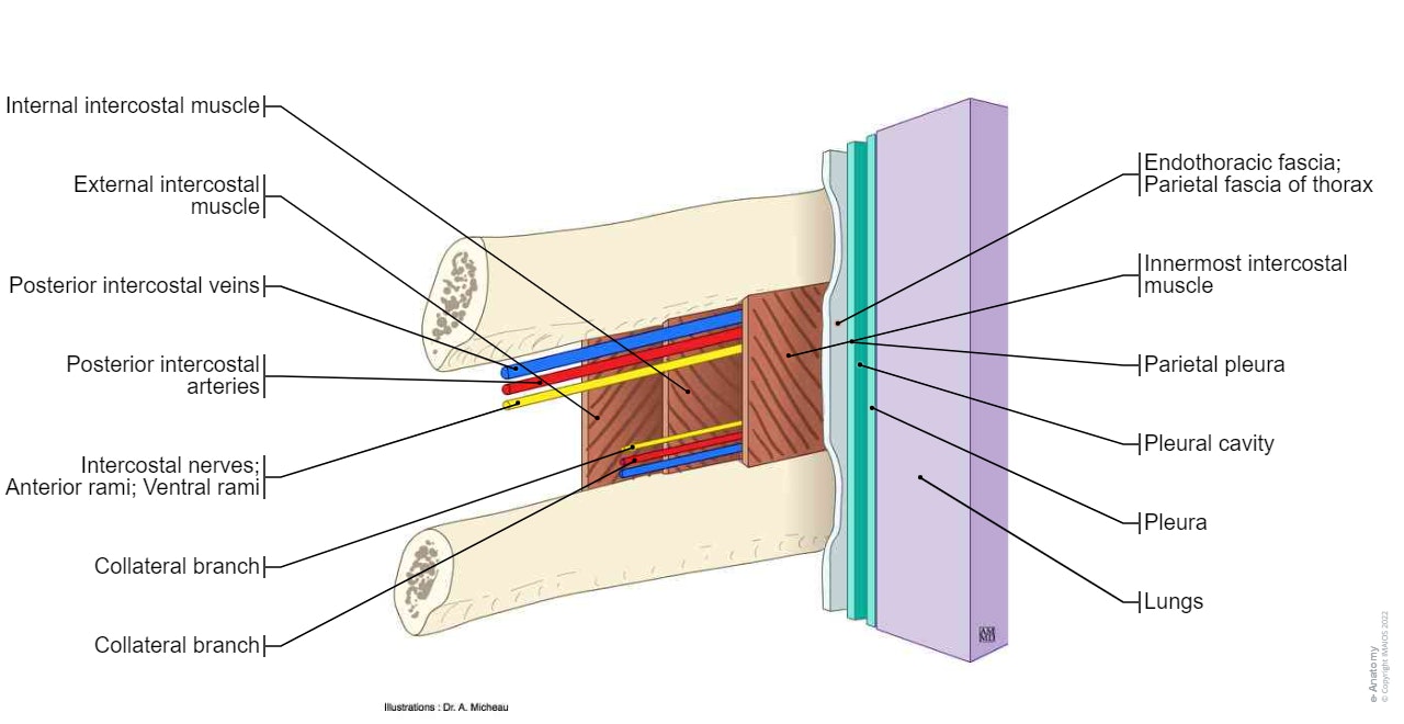
  • Chest muscles: diagrams showing the arrangement of the chest muscles (pectoralis major and minor muscles, serratus anterior and intercostal muscles...)


Muscles of thorax : Sternalis; Sternalis muscle, Pectoralis major, Pectoralis minor, Subclavius, Serratus anterior


Intercostal space - Anatomy diagram : External intercostal muscle, Internal intercostal muscle, Innermost intercostal muscle, Endothoracic fascia; Parietal fascia of thorax, Posterior intercostal arteries, Posterior intercostal veins, Intercostal nerves; Anterior rami; Ventral rami


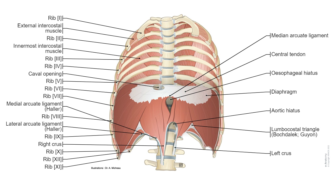
  •  Diaphragm: different views illustrating the different anatomical parts of the diaphragm (pillars, central tendon, arcuate ligament...)


Diaphragm : Diaphragm Lumbar part of diaphragm Right crus Left crus Median arcuate ligament Medial arcuate ligament Lateral arcuate ligament Costal part of diaphragm Sternal part of diaphragm Aortic hiatus Oesophageal hiatus Phrenicooesophageal ligament Central tendon Caval opening Sternocostal triangle Lumbocostal triangle


  • Arteries: represents the anatomy of the arterial vascularisation of the thoracic cage (intercostal arteries, phrenic arteries, internal mammary arteries)
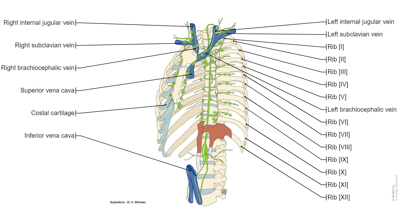
  • Veins: diagrams of intercostal veins, internal mammaries, the azygos vein and the phrenic veins.
  • Thoracic nerves: diagram of spinal thoracic nerve divided into the intercostal nerve and its vertebral and mammary branches.
  • Thoracic lymph nodes: anatomy of the thoracic duct, the cisterna chyli, and parietal thoracic lymphatic ganglia (parasternal and intercostal nodes...)


Thoracic duct: Arch of thoracic duct, Cisterna chyli; Chyle cistern, Thoracic lymph nodes, Parasternal nodes, Intercostal nodes, Superior diaphragmatic nodes


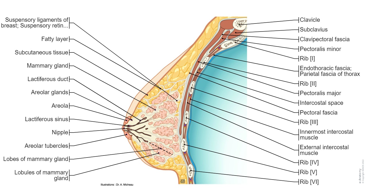
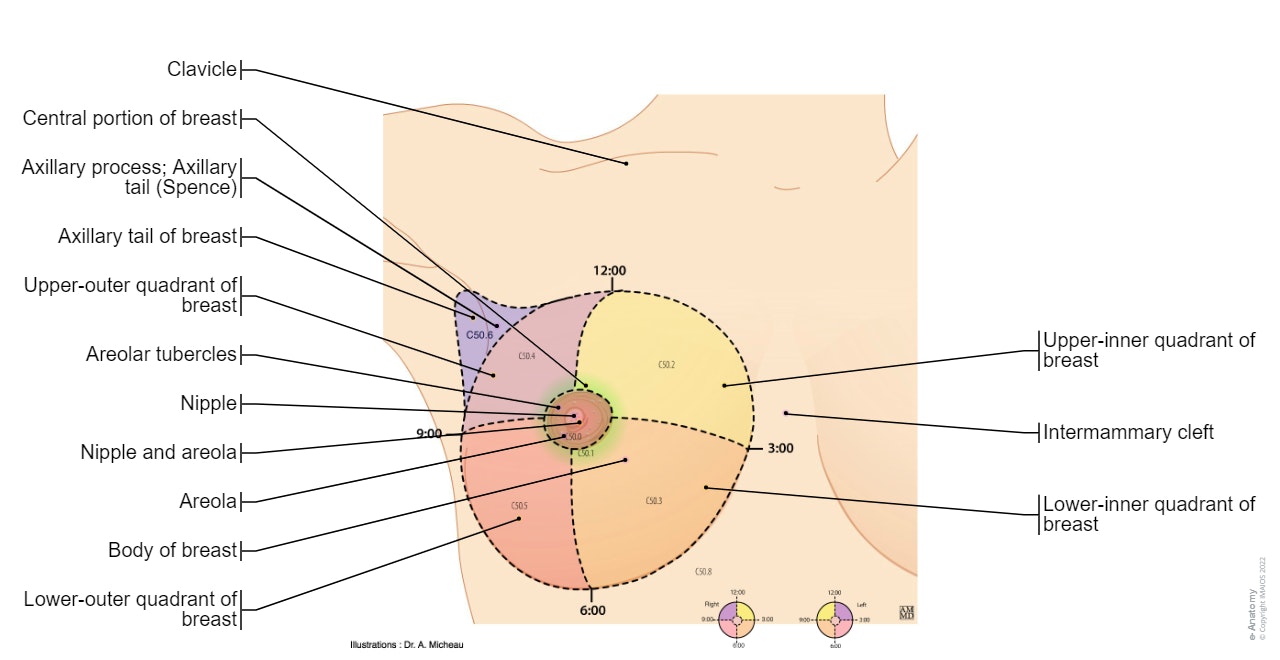
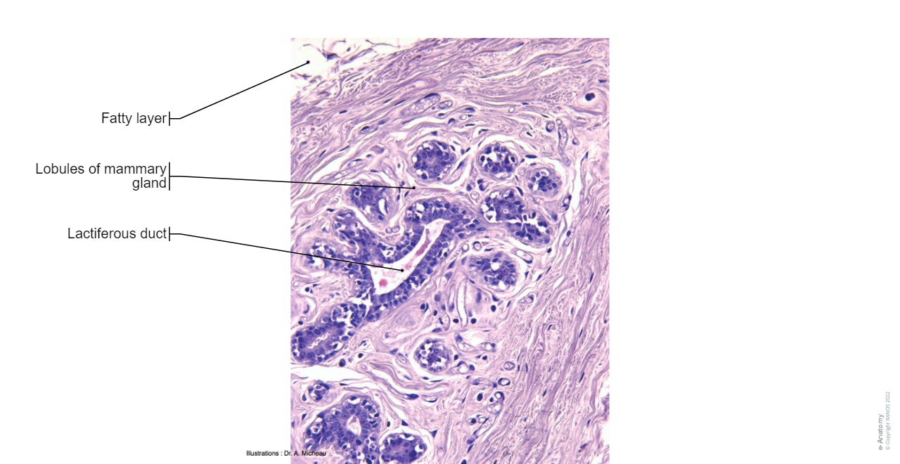
  •  Breast: several illustrations represent the surface anatomy of the mammary gland (nipple, areola, axillary process), the sagittal anatomy of the breast (lobes, lactiferous sinuses, milk ducts), the arterial and venous vascularisation, the lymphatic drainage and histology of the breast.


Quadrants of the breast: Nipple and areola, Central portion of breast, Axillary tail of breast


Breast - Histology : Mammary gland,Lobes of mammary gland, Lobules of mammary gland, Lactiferous duct, Lactiferous sinus


Labels of the anatomical structures of the thoracic wall and breast:

260 anatomical structures were labeled on this atlas of thoracic anatomy, accessible via the "anatomical structures" tab:

  • Regions
  • Plans and lines
  • Bones: Ribs, Sternum, Thoracic wall, Spine, Bones of the upper limb
  • Joints
  • Ligaments
  • Muscles
  • Diaphragm
  • Arteries
  • Veins
  • Lymph nodes
  • Nerves
  • Breast
  • Other structures


Language and terminology for the study of the anatomy of the thorax

The anatomical structures are available in different languages (English, French, Japanese, German, Chinese, Portuguese, Russian, Polish, Italian, Korean and Spanish) adapted from the Terminologia Anatomica.

Breast - Anatomy : Sagittal section (Illustrations: A. Micheau - MD - Imaios)
Breast
Trunk - Surface anatomy : Pectoral region, Mammary region, Hypochondrium, Presternal region, Midclavicular line
Trunk
Anatomy of the thoracic wall and the breast (Illustrations) : Thoracic cage - Thoracic cavity
Anatomy of the thoracic wall and the breast (Illustrations) : Thoracic cage
Sternum (Muscles-Attachment) : Manubrium of sternum Clavicular notch Jugular notch; Suprasternal notch Sternal angle Body of sternum Xiphoid process Costal notches
Sternum (Muscles
Muscles of thorax : Sternalis; Sternalis muscle, Pectoralis major, Pectoralis minor, Subclavius, Serratus anterior
Intercostal space - Anatomy diagram : External intercostal muscle, Internal intercostal muscle, Innermost intercostal muscle, Endothoracic fascia; Parietal fascia of thorax, Posterior intercostal arteries, Posterior intercostal veins, Intercostal nerves; Anterior rami; Ventral rami
Intercostal space
Diaphragm : Diaphragm Lumbar part of diaphragm Right crus Left crus Median arcuate ligament Medial arcuate ligament Lateral arcuate ligament Costal part of diaphragm Sternal part of diaphragm Aortic hiatus Oesophageal hiatus Phrenicooesophageal ligament Central tendon Caval opening Sternocostal triangle Lumbocostal triangle
Thoracic duct: Arch of thoracic duct, Cisterna chyli; Chyle cistern, Thoracic lymph nodes, Parasternal nodes, Intercostal nodes, Superior diaphragmatic nodes
Quadrants of the breast: Nipple and areola, Central portion of breast, Axillary tail of breast
Breast - Histology : Mammary gland,Lobes of mammary gland, Lobules of mammary gland, Lactiferous duct, Lactiferous sinus
Breast
  • Terminologia Anatomica:International Anatomical Terminology - FCAT Federative Committee On Anatomical Terminology, Federative Committee on Anatomical Terminology - Thieme, 1998 - ISBN 3131152516, 9783131152510
  • Atlas d'anatomie humaine- 4e édition - Frank-H Netter - Pierre Kamina (Traducteur) - Paru le : 25/07/2007 - Editeur : Masson - ISBN : 978-2-294-08042-5 - EAN : 9782294080425  (lien : http://www.netterimages.com/)
  • Lexique illustré d'anatomie Feneis– Feneis - Editeur : Flammarion Médecine-Sciences (23 août 2007)  - Collection : ATLAS DE POCHE - ISBN-10: 225712250X - ISBN-13: 978-2257122506
  • Gray's Anatomie pour les étudiants- de Richard-L Drake, Wayne Vogl et Adam-W-M Mitchell - Editeur : Elsevier (24 octobre 2006) - Langue : Français - ISBN-10: 2842997743 - ISBN-13: 978-2842997748